- The exact cause of Parkinson’s disease remains unknown; however, researchers know some genetic factors contribute to the condition, such as a mutation in the PINK1 gene.
- A new study for the first time explains what human PINK1 looks like and how it is activated.
- Researchers believe these findings may help one day lead to new treatment options for Parkinson’s disease.
Researchers estimate that about 10 million people globally live with Parkinson’s disease — a neurological condition that affects movement.
While the exact cause of Parkinson’s disease is still unknown, past studies show that a combination of both environmental and genetic factors contribute to the condition.
For instance, scientists have discovered that having certain genes may increase a person’s risk for developing Parkinson’s disease, such as a mutation in the PINK1 (PTEN-induced putative kinase 1) gene.
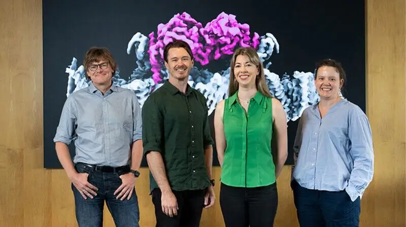
Now, a new study recently published in the journal Science for the first time explains what human PINK1 looks like and how it is activated.
Researchers believe these findings may help one day lead to new treatment options for Parkinson’s disease.
PINK1 plays an important role in the body as the protein it expresses helps protect mitochondria — the cellular energy sources — from harm, and leads their removal from the body when they do become damaged over time.
“[The] PINK1 [protein] acts as a beacon for damaged mitochondria, famously known as the ‘powerhouse of the cell’,” Sylvie Callegari, PhD, senior research officer in the Ubiquitin Signalling Division at The Walter and Eliza Hall Institute of Medical Research in Australia, and both first and corresponding author of this study told Medical News Today.
“PINK1 senses this damage and docks to the surface of mitochondria. Once positioned on the mitochondrial surface, PINK1 becomes active and seeks out a small protein known as
“When damaged mitochondria are not cleaned up, they release toxins, which kill cells,” she continued. “Brain cells, such as neurons, which require a lot of energy and have a lot of mitochondria are particularly sensitive to mitochondrial toxicity and are more likely to die. The death of neuronal cells in the brain is what causes Parkinson’s disease.”
Callegari said that up until now, one of the main obstacles that has prevented researchers from seeing what PINK1 looks like was that there is not very much PINK1 in the cell.
“Until now, scientists had been studying insect PINK1 because it is possible to produce it in large amounts, and by visualizing insect PINK1, we were able to decipher how it can be activated,” she explained.
“However, we were never able to see how PINK1 docks to the mitochondrial surface, which is the important step that precedes its activation. To get around this problem, we used very large amounts of cells (nearly 10L) to get enough human PINK1 that we could then use for visualization using
Callegari and her team discovered there are four main steps to how PINK1 works: Sensing mitochondrial damage, attaching to the damaged mitochondria, tagging ubiquitin, and then ubiquitin links to a protein called Parkin to “recycle” the injured mitochondria.
“PINK1 is special because it can alter the ubiquitin tag by placing an additional marker on it — it basically tags the tag,” Callegari said. “This alteration is a very specific signal that initiates the disposal of the entire mitochondria.”
How PINK1 causes Parkinson’s“When PINK1 is mutated, it cannot perform its signaling function, and so mitochondria are not effectively cleaned up. Defective mitochondria are toxic to cells, resulting in cell death. The death of neuronal cells in the brain causes Parkinson’s disease.”
— Sylvie Callegari, PhD
Callegari believes their findings bring us a big leap closer to developing therapies for Parkinson’s disease.
“Ideally, we want to design a drug that makes PINK1 more active, but without the ability to see PINK1, it is very hard to develop a drug to do this,” she explained. “Now that we can see PINK1, we have the blueprint that we need to improve its activity.”
“There are currently some drugs in the pipeline that are believed to increase the activity of PINK1, but without ever seeing where or how these drugs interact with PINK1, we don’t have a complete understanding of how they work. We plan to use our approach to visualize these drugs in association with PINK1 to understand how they work. Furthermore, we will also use our PINK1 model to design new drugs that increase PINK1 activity.”
— Sylvie Callegari, PhD
MNT spoke with Daniel Truong, MD, neurologist and medical director of the Truong Neuroscience Institute at MemorialCare Orange Coast Medical Center in Fountain Valley, CA, and editor-in-chief of the Journal of Clinical Parkinsonism and Related Disorders, about this study.
“As a physician who treats patients with Parkinson’s disease, the recent elucidation of the human PINK1 protein structure bound to mitochondria is a significant and encouraging development,” Truong commented.
“Mutations in the PINK1 gene have been linked to early-onset Parkinson’s disease. By resolving the structure of PINK1, researchers have provided deeper insights into its function and how its dysfunction can lead to neurodegeneration,” he added.
“Understanding the structural configuration of PINK1 will open avenues for developing targeted therapies aimed at modulating its activity. This could lead to interventions that enhance mitochondrial quality control mechanisms, potentially slowing or halting disease progression. With the understanding of the structural blueprint of PINK1, pharmaceutical research can focus on designing molecules that interact precisely with this protein leading to more effective treatments with fewer side effects.”
— Daniel Truong, MD
MNT also spoke with two neurologists from Hackensack Meridian in New Jersey about this research.
Rocco DiPaola, MD, neurologist and movement disorder specialist at Hackensack Meridian Neuroscience Institute at the Jersey Shore University Medical Center commented that this study is another positive step in uncovering and understanding the mechanisms involved in hereditary forms of Parkinsonism.
“It is important to continue to look for new ways to treat Parkinson’s disease, as current therapies have their limitations, primarily addressing
Umer Akbar, MD, neurologist and director of the Movement Disorder Center in the Department of Neurology and Hackensack Meridian Neuroscience Institute at Hackensack University Medical Center, said that while this discovery provides crucial foundational knowledge, but translating this knowledge to find a cure or an effective treatment will require further research.
“Drug development is a long and complex process, and many promising drug candidates fail in clinical trials. However, this discovery undoubtedly removes a major obstacle and significantly increases the chances of developing effective therapies for Parkinson’s disease in the future. It provides a much-needed roadmap for future research and drug development efforts.”
— Umer Akbar, MD