- Researchers designed a wearable device that can be used to screen for breast cancer at home.
- Initial tests show it can detect small cysts similar in size to early-stage breast tumors.
- Clinical trials are needed to verify the device’s efficacy.
A novel, wearable technology that can be attached to a bra may one day help people detect signs of breast cancer from the comfort of their homes.
Breast cancer is the most prevalent cancer
Breast cancer has a 5-year relative
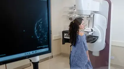
Currently, an X-ray imaging method known as a mammogram is the most commonly used technique for screening breast cancer.
While mammograms are generally effective—detecting around
Efforts to improve accessibility and reduce costs of breast cancer screening could improve health outcomes for people diagnosed with the condition worldwide.
Recently, researchers designed a wearable ultrasound breast patch that could help patients screen for breast cancer from their homes.
Dr. Kamila Seilhan, a board certified physician and Chief Medical Director with LabFinder.com, not involved in the research, told Medical News Today:
“This is a wearable ultrasound device that could allow people to detect tumors early and help patients at high risk of developing breast cancer in between routine mammograms.”
“A wearable ultrasound breast patch makes it possible to get uniform and repeatable images of the whole breast without relying on special operator training. […] The patch has an easy-to-use tracker that allows for large-area, deep scans, and images from multiple angles of the breast. It is a safe way to track changes in soft tissue in real time,” she added.
The research was published in Science Advances.
The device is based on the same ultrasound technology used in imaging centers. However, its piezoelectric-based materials allow it to be miniaturized into a portable ultrasound scanner.
“The device sends sound waves into the breast tissue, and as it moves across the breast it produces high-quality images identifying cysts that may need to be investigated by a breast cancer specialist,” Dr. Jennifer Tseng, F.A.C.S., medical director of breast surgery at City of Hope Orange County and a double board-certified surgical oncologist specializing in breast cancer at City of Hope Orange County Lennar Foundation Cancer Center in Irvine, California, not involved in the research, told MNT.
The researchers designed a flexible, 3D-printed patch with honeycomb-like openings to make the device wearable. The patch attaches to a bra with openings that allow it to touch the skin, where it can scan breast tissue. The scanner can be placed in six different positions, allowing the entire breast to be imaged. It can also be rotated to take images from different angles.
Already, the researchers have tested the scanner on a 71-year-old woman with a history of breast cysts. Using the device, they could detect cysts as small as 0.3 centimeters in diameter- the size of early-stage tumors. They reported that the resulting images had a similar resolution to traditional ultrasounds and a depth of roughly 80mm.
Canan Dagdeviren, Ph.D., Associate Professor of Media Arts and Science at Massachusetts Institute of Technology (MIT), senior author of the study, told MNT that the device makes it easy to capture images from the same position time and time again. This makes it ideal for long-term monitoring, especially as ultrasounds do not pose radiation risk, unlike mammograms,
Dr. Dagdeviren noted that her ultimate goal with the device is to make screening for breast cancer more affordable and reach underrepresented women- including those in less economically-developed countries.
If proven effective, Dr. Seilhan noted that the device could be particularly useful in remote areas without easy access to medical centers.
“The device’s low cost makes it easier for Healthcare facilities and organizations with limited funds to buy it,” she said.
She added that as the device is easy to use, it could also be helpful in places where medical workers have limited levels of technical knowledge.
Dr. Tseng noted, however, that while greater access to diagnostic technologies is crucial for patients in less-developed countries, specialized expertise is also important to benefit from them fully.
“While this device could help patients detect potential trouble spots that they could not detect before, they still need to have the data reviewed by an expert who can recommend what to do next,” she said.
Dr. Dagdeviren told MNT that the device may become available for use within 4-5 years. To this end, she is launching a company and looking for investors and partners.
“We need around $40M to get FDA approval and do mass production,” she said.
She added that while the device currently requires a ‘bulky computer interface’ to process images, her team is working on a more compact form and will publish an iPhone-size image processor soon.
The researchers are also developing a workflow that will allow artificial intelligence to analyze data and generate diagnostic assessments that may be more accurate than those performed by a radiologist comparing images taken years apart.
To understand more about what may lie ahead for the device, MNT spoke with Dr. Richard Reitherman, Ph.D., board certified radiologist and medical director of breast imaging at MemorialCare Breast Center at Orange Coast Medical Center in Fountain Valley, CA, not involved in the study.
“If this kind of product can be demonstrated to be on par with mammography and dedicated breast ultrasound for breast cancer screening, it will be a welcomed supplemental addition to women’s health care,” he said.
He noted, however, that successful clinical trials are one of the major challenges for any new devices and that these will likely need to be undertaken alongside the American College of Radiology.
“This is a complex and difficult proposition,” he noted, “The jump from translational science to clinical efficacy remains to be seen.”
The device is still in the early developmental stage and thus has limitations.
Dr. Onalisa Winblad, a breast radiologist at The University of Kansas Cancer Center, not involved in the study, told MNT that she does not currently endorse its use as it “does not have scientific data to prove utility.”
“The images provided in the linked article are of poor quality [in comparison] to our standard breast ultrasound images. Additionally, ultrasound is a tool that is helpful in conjunction with mammography in patients with dense breast tissue.”
Dr. Tseng agreed that while ultrasound is a valuable tool for screening for breast cancer, it cannot replace mammograms and other preventive care from a breast cancer expert.
“Different technologies detect different types of breast changes better than others. For example, some calcifications can be detected through mammography but not through ultrasound,” she said.
Dr. Seilhan acknowledged that before widespread use, the scanner should be carefully tested to see how well it detects problems within breasts.
“The device’s ability to find true positive cases and avoid fake positive cases are two of the most important factors in how well it works as a breast cancer screening tool,” she noted.
She added that although the device might be easy to use, its efficiency could still depend on how skilled the person using it is. She further noted that large-area and deep-tissue imaging can be difficult as the human breast varies between people and even in the same person over time.
When asked about the device’s limitations, Dr. Reitherman noted that the scanner must be used under medical supervision- such as through ‘virtual supervision by a radiologist- to maintain proper quality metrics.
“Therefore, the existing medical community and physicians that would be interpreting and recommending actions based on this device’s information would need to be on board,” he noted.
“Patient wearable, self-monitoring remote devices currently in use for cardiac and diabetic applications require a well-developed and sophisticated infrastructure. This involves many steps and personnel that will interpret, analyze, and initiate actions based on remote data input. It is this integrative process that will require significant participation,” he concluded.


